
SPRm 200 System is the world’s first commercial Surface Plasmon Resonance Microscopy (SPRM). It combines the high spatial resolution of optical microscopy with the powerful sensing capability of SPR, making it possible to measure the binding of molecules label free with membrane proteins on single intact cells for the first time. Cells are incubated on a traditional SPR sensor surface, the analyte molecules are introduced with a flow system to allow the binding of the molecules with membrane receptors on the cells, and the binding kinetics is obtained for each of the cells on the surface from the single-cell sensorgrams. The association constant (ka), dissociation constant (kd), as well as the equilibrium constant (KD) are obtained. Unlike traditional SPR systems, SPRm 200 provides high spatial resolution and simultaneous optical microscopy capability, which are necessary to resolve and measure molecular binding taking place on the surface of individual cells. With SPRm 200, the SPR response is mapped in a 600 µm x 450 µm image and the binding affinity and kinetics information is collected at each pixel of the image. By providing a sensorgram at each pixel of the image, the instrument offers spatial mapping of the biding activities over any given sensing area, yielding much more information than the conventional SPR systems.
A particularly unique application of SPRM for studies of biomolecular interactions with multiple cells is exemplified by the studies of binding between glycoproteins at cell membranes and their ligands. Glycoproteins, which are important for cell recognition and communications, are membrane proteins with sugar groups extended into the extracellular space of the cells (1). The interaction of wheat germ agglutinin (WGA, 36k MW), a lectin that binds sugar groups and can recognize N-acetylglucosamine (GlcNAc), on live and fixed Barrett’s esophagus-derived CP-D (CP-18821) cells was investigated in this note. WGA exposure to these cells resulted in an increase of SPR response in the SPR image, which indicates the presence of sugar residues on the glycan chain of the membrane protein (2).
Fig.1 shows the application of SPRm 200 to measure the binding of lectin with glycoproteins on the membranes of individual live cells. The bright-field optical microscopy of SPRm 200 reveals the individual cells (Fig. 1A), while the SPRM image (Fig. 1B) provides valuable binding signal on each of the cells at a specific concentration, from which SPR sensorgrams for the individual cells are obtained (Fig. 1C) for wheat germ agglutinin (WGA). For this study, the cells were incubated on the sensor surface at 37°C with 5% CO2 and 70% RH. The cell surface was regenerated with 50 mM N-acetylglucosamine (GlcNAc). Only two concentrations (87nM & 175nM) were tested in this set up due to cell apoptosis.
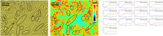
Fig 1. Bright field image of live CP-D (A), SPR image of CP-D binding to glycoprotein receptors (B), SPR sensorgrams of selected cells (C). From the eleven regions (live cells) selected for analysis, the average ka, kd and KD were 1.14 x10⁴ M-1 s-1, 3.42 x 10⁻⁴ s-1 and 38.7 (+/-24) nM, respectively.
Figure 2 shows the binding results of 20 fixed cell regions. In addition to the regeneration and conditions described above, the cells were fixed with 4% of PFA (paraformaldehyde) for 30 min at room temperature. Five concentrations (43nM to 0.7uM) of WGA were introduced to the system to test the binding to the cells. The average ka, kd and KD were 1.54 x10⁴ M-1 s-1, 3.34 x10⁻⁴ s-1 and 20.1 (+/- 6.6) nM respectively.
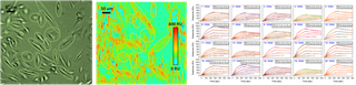
Fig 2. Bright field image of fixed CP-D (A), SPR image of CP-D binding to glycoprotein receptors (B), SPR sensorgrams of selected cells (C).
In conclusion, SPRM is a powerful technique that is useful to study the kinetic behavior of live or fixed cells in vitro without the need of labels or extracting/purifying the membrane from its natural condition. With SPRm 200, visualization of the cells can be achieved using the optical microscope, the binding behavior can be observed via the SPR image and the ka, kd, and KD can be obtained from any given number of sensorgrams selected from the regions of interest.
DOWNLOAD PDF
Download a PDF of Application Note 124: Quantifying Molecular binding to Membrane Proteins on Individual Cells with Surface Plasmon Resonance Microscopy
- Dell, A. et al, Science, 2001, 291, 2351-2356
- Wang, W. et al, Nature Chemistry, 2012, 4, 846–853